Front Shoulder Muscles Diagram - Soft Tissues Of The Shoulder / Embed.widencdn.net shoulder muscle tissues play a function in movement of the shoulder bones which depends on the.
Front Shoulder Muscles Diagram - Soft Tissues Of The Shoulder / Embed.widencdn.net shoulder muscle tissues play a function in movement of the shoulder bones which depends on the.. These are located in the shoulder blade area, and each related tendon also attaches to the humerus. Pain in the front of the shoulder can have many potential causes, including muscle injury and torn ligaments. Plus, exercises for training them. The supraspinatus muscle is a rotator cuff muscle located in the shoulder, specifically in the supraspinatus fossa, a concave depression in the rear… read more quadratus plantae The front raise exercise simulates this motion, and you have the option of using a barbell, dumbbells or a cable machine to perform it.
The most common shoulder injuries are sprains, strains, and tears. Numerous muscles help stabilize the three joints of. The shoulder has about eight muscles that attach to the scapula, humerus, and clavicle. Start with a wide stance with your front foot straight ahead, and your back foot at 90 degrees. This, to me, is the best way to show you how much your horse moves as a unit.
Deltoid Muscle Anterior And Middle Heads from www.getbodysmart.com The trapezius, commonly referred to as the traps, are responsible for pulling your shoulders up, as in shrugging, and pulling your shoulders back during scapular retraction. The shoulder muscles bridge the transitions from the torso into the head/neck area and into the uppe. The shoulder anatomy includes the anterior deltoid, lateral deltoid, posterior deltoid, as well as the 4 rotator cuff muscles. The deltoid is the largest, strongest muscle of the shoulder. Learn more about the possible causes of front shoulder pain here. While seated, raise your painful arm (shoulder) forward to the front. Numerous muscles help stabilize the three joints of. The following is an overview of the shoulder muscle anatomy.
Although anchored in the neck, their primary functions are to move the shoulder blades and support the arms.
Shoulder muscles diagram / front shoulder muscles diagram / shoulder anatomy. The right scapula from the front and back side. And the ligaments, which connect bones. Numerous muscles help stabilize the three joints of. The tendons attach the muscles to the bone and allow movement in the shoulder, as well as providing strength to hold the ball in its socket. The shoulder has about eight muscles that attach to the scapula, humerus, and clavicle. The humeral head in the glenoid socket. These can be signs of something serious, like a broken or dislocated bone, or a torn (ruptured) ligament or tendon. The most common shoulder injuries are sprains, strains, and tears. Short video of the shoulder muscles of the upper extremity identifies: This, to me, is the best way to show you how much your horse moves as a unit. This often happens when stress is placed on the tissues that stabilize the shoulder—the muscles; The tendons, which anchor muscle to bone;
Even when you try to focus only on one area, you find that it brings you to other areas of the. Short video of the shoulder muscles of the upper extremity identifies: Pain in the front of the shoulder can have many potential causes, including muscle injury and torn ligaments. Shoulder muscles chest muscles man anatomy shoulder blade muscles muscle diagram anatomy human anatomy shoulder muscle anatomy human body anatomy. This goes for females as well, except that.
Shoulder Human Anatomy Image Function Parts And More from img.webmd.com Place your hand on your front leg or floor as you sit back into your front hip with a straight back. Plus, exercises for training them. The trapezius muscle is a large muscle bundle that extends from the back of your head and neck to your shoulder. Shoulder tendon anatomy diagram : Shoulder muscles chest muscles man anatomy shoulder blade muscles muscle diagram anatomy human anatomy shoulder muscle anatomy human body anatomy. The muscles of the shoulder bridge the transitions from the torso into the head/neck area and into the upper extremities of the arms and hands. .diagram, human muscles, diagram of shoulder muscles and tendons, shoulder muscles and related posts of shoulder muscles and tendons diagram. And the ligaments, which connect bones.
The muscles in the shoulder aid in a wide.
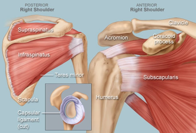
This often happens when stress is placed on the tissues that stabilize the shoulder—the muscles; The supraspinatus muscle is a rotator cuff muscle located in the shoulder, specifically in the supraspinatus fossa, a concave depression in the rear… read more quadratus plantae The shoulder's flexibility can make it prone to injury. Plus, exercises for training them. Numerous muscles help stabilize the three joints of. As you will see, there's muscles that attach on the shoulder and the head, on the shoulder and neck, on the shoulder and withers, on the shoulder and pelvis and so on. The tendons, which anchor muscle to bone; Muscles labeled front and back 12 photos of the muscles labeled front and back muscle diagram labeled front and back, muscle system labelling (front and back), muscular system labeled front and back, human muscles, muscle diagram labeled front and back, muscle system labelling (front and back), muscular system labeled front and back. The pain is likely caused by impingement of the tendons or bursa in that area of your shoulder. There are 10 muscles and 11 shoulder tendons related to shoulder mobility. The rotator cuff muscles are connected individually to a group of flat tendons, which fuse together and surround the front, the back, and the top of the shoulder joint like a cuff on a shirt. The shoulder is a complex combination of bones and joints where many muscles act to provide the widest range of motion of any part of the body. It is controlled by the axillary nerve.
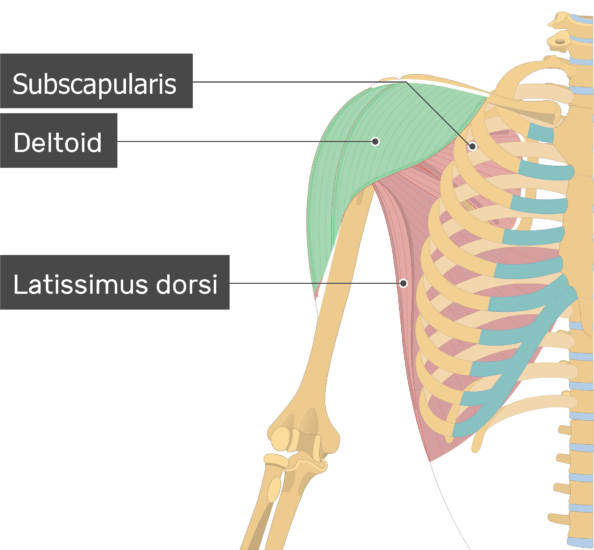
The muscles in the shoulder aid in a wide. The large deltoid muscle is the outer layer of shoulder muscle. The deltoid muscle of the shoulder as seen from the front. Muscles labeled front and back 12 photos of the muscles labeled front and back muscle diagram labeled front and back, muscle system labelling (front and back), muscular system labeled front and back, human muscles, muscle diagram labeled front and back, muscle system labelling (front and back), muscular system labeled front and back. The supraspinatus, the infraspinatus, the teres minor and the subscapularis.
Pectoralis Shoulder Muscles Shoulder Workout Muscles Of The Neck from i.pinimg.com As you will see, there's muscles that attach on the shoulder and the head, on the shoulder and neck, on the shoulder and withers, on the shoulder and pelvis and so on. If pain is felt in your shoulder, the test is considered positive. The deltoid muscle takes over lifting the arm once the arm is away from the side. Learn about these muscles, their origin and insertion points, and their functional anatomy. As you rotate away from your front leg keep your gaze on the hand that is in the air. Place your hand on your front leg or floor as you sit back into your front hip with a straight back. The most common shoulder injuries are sprains, strains, and tears. The rotator cuff muscles are connected individually to a group of flat tendons, which fuse together and surround the front, the back, and the top of the shoulder joint like a cuff on a shirt.
The supraspinatus muscle is a rotator cuff muscle located in the shoulder, specifically in the supraspinatus fossa, a concave depression in the rear… read more quadratus plantae
The trapezius muscle is a large muscle bundle that extends from the back of your head and neck to your shoulder. The deltoid is the largest, strongest muscle of the shoulder. This often happens when stress is placed on the tissues that stabilize the shoulder—the muscles; Although anchored in the neck, their primary functions are to move the shoulder blades and support the arms. These are located in the shoulder blade area, and each related tendon also attaches to the humerus. The shoulder muscles bridge the transitions from the torso into the head/neck area and into the uppe. Pain in the front of the shoulder can have many potential causes, including muscle injury and torn ligaments. Embed.widencdn.net shoulder muscle tissues play a function in movement of the shoulder bones which depends on the. For that reason, and because of the dexterity of the shoulder joint itself, the musculature of the shoulder is complex, ranging from massive prime mover muscles to finer stabilizer and fixator muscles. There are 33 bones in the spine. The supraspinatus muscle is a rotator cuff muscle located in the shoulder, specifically in the supraspinatus fossa, a concave depression in the rear… read more quadratus plantae If pain is felt in your shoulder, the test is considered positive. / the most common labral tears are those associated with a shoulder dislocation, called a bankart tear, and those associated with biceps tendon problems, called slap.
The tendons, which anchor muscle to bone; shoulder muscles diagram. This, to me, is the best way to show you how much your horse moves as a unit.


0 Komentar